
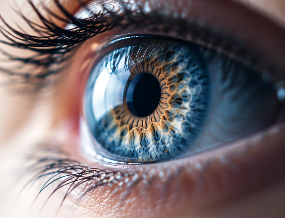
What the cornea does
Light enters the eye through the cornea first. Because it is curved, it bends that light and provides most of the eye’s focusing power. It must stay perfectly clear and smooth to give sharp vision.
Diseases of the cornea and the eye’s surface (the “external” eye) are treated by cornea specialists.
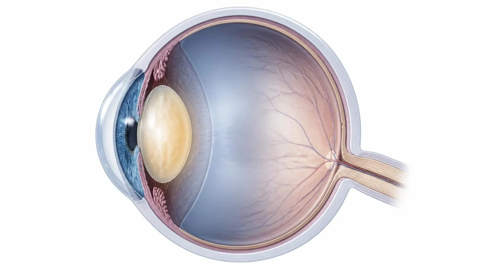
Five thin layers
The cornea is only about half a millimeter thick, yet it has five layers. The outer surface protects the eye, the middle gives it strength and clarity, and a single inner layer of cells constantly pumps fluid out so the cornea stays clear.
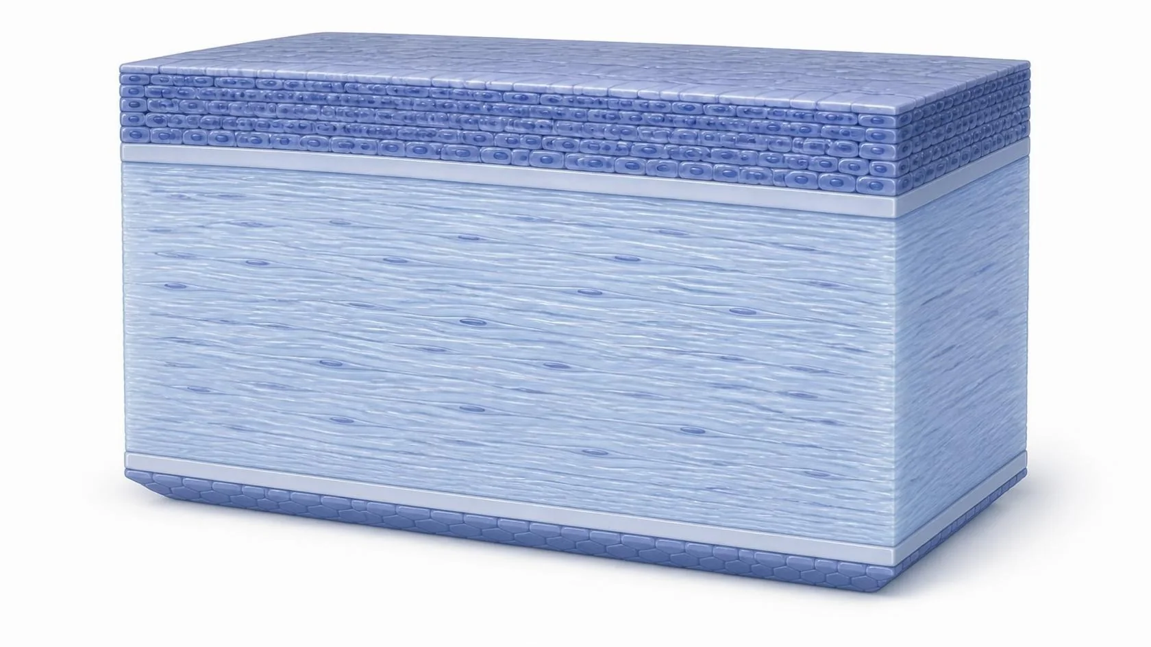

 Healthy vision With corneal disease
Common corneal conditions
Dry eye & ocular surface disease
An unstable tear film or irritated surface can cause burning, grittiness and fluctuating vision.
Keratoconus
The cornea gradually thins and bulges into a cone shape, distorting vision. It often begins in the teens or twenties.
Fuchs’ dystrophy
The inner pumping cells slowly fail, so the cornea swells. Vision is often worst in the morning.
Scars & infections
Injuries and infections such as corneal ulcers can leave the cornea cloudy or irregular.
Growths on the surface
Growths such as a pterygium can spread onto the cornea and affect comfort and vision.
Healing problems
Some eyes struggle to heal the surface layer, which leaves the cornea vulnerable.
How it can be treated
- 1
Medical care
Lubricating and prescription drops, ointments and treatment of the eyelids and tear film.
- 2
Specialty contact lenses
Custom lenses can smooth an irregular cornea, such as in keratoconus, and sharpen vision.
- 3
Corneal crosslinking
Special drops and ultraviolet light strengthen the cornea to help slow or stop keratoconus from progressing.
- 4
Corneal transplant
Replacing damaged tissue with healthy donor tissue. Today, often only the affected layers are replaced (DMEK, DSAEK or DALK) instead of the full thickness (PK).
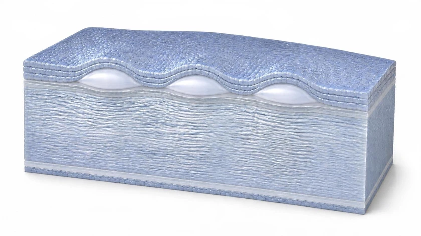 Also offered Superficial keratectomy (SK) Smoothing the front surface of the cornea for Salzmann’s nodules, ABMD and band keratopathy. Learn about SK
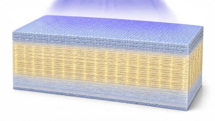 Also offered Keratoconus & crosslinking Strengthening a thinning, cone-shaped cornea to help stop keratoconus from progressing. Learn about crosslinking
See a doctor promptly if you notice
- A red, painful eye, especially if you wear contact lenses
- Sudden blurred vision or sensitivity to light
- A white or gray spot on the colored part of the eye