

How DMEK is performed
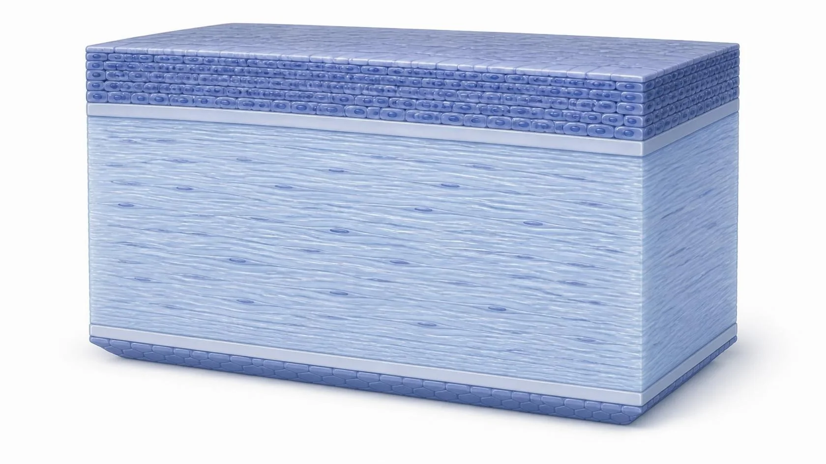
Think of the cornea as a wall and its inner layer as the wallpaper. In Fuchs’ dystrophy and similar conditions, the wall is still sound, but the wallpaper has failed. That inner layer of cells normally pumps fluid out so the cornea stays clear; when it fails, the cornea swells and vision turns cloudy.
DMEK simply replaces the wallpaper. The damaged inner layer is gently removed, and a healthy one from a donor cornea is rolled up, slipped in through a small opening, unfolded into place and held there with a bubble of air while it attaches. The rest of your own cornea stays untouched.
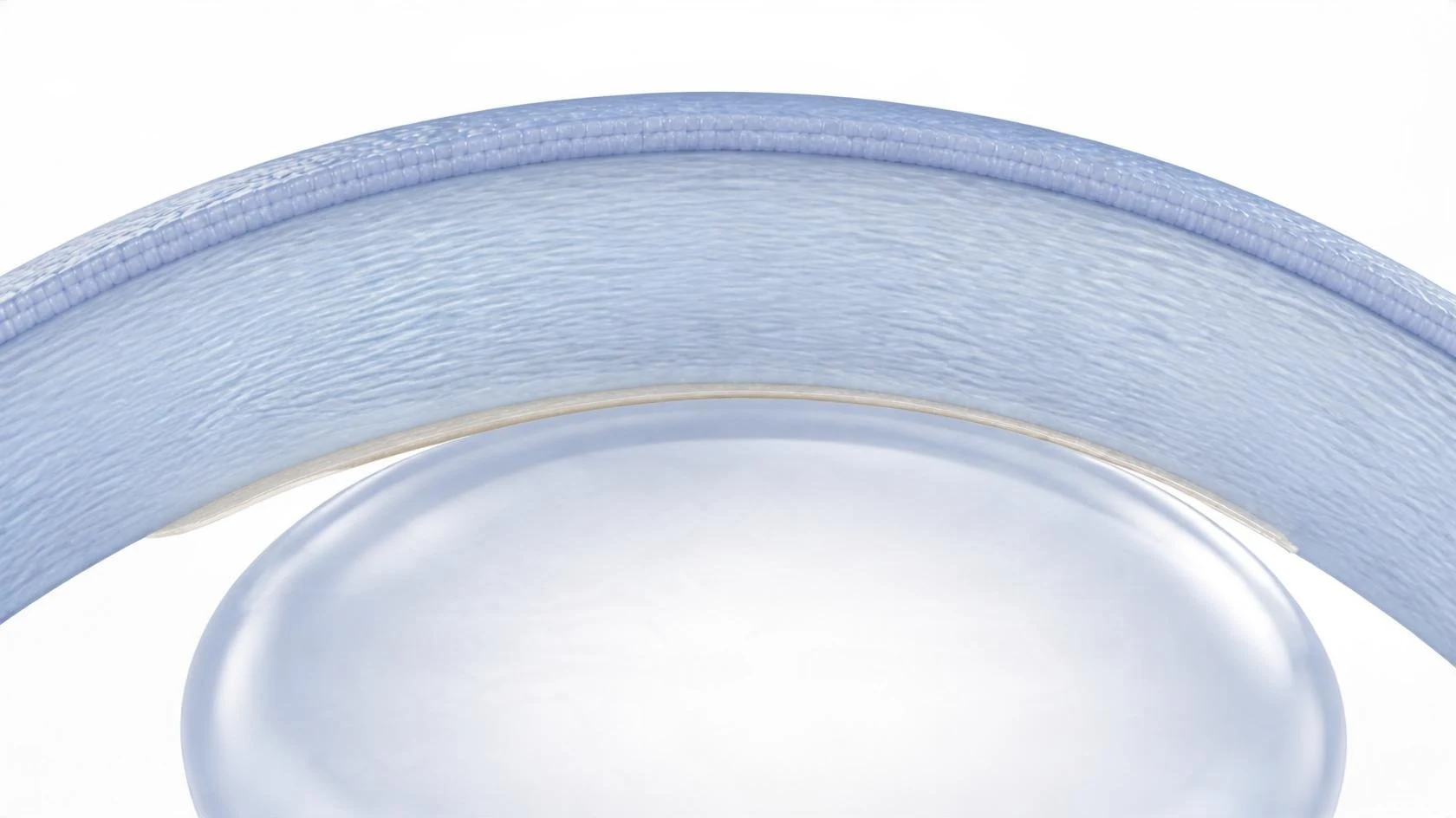
Placing the new layer
The donor layer is thinner than a human hair and naturally curls into a scroll. Your surgeon guides it through the small opening, gently unrolls it against the back of your cornea, then adds a bubble of air to hold it in place while it seals on.
Over the following days the bubble is absorbed. As the new cells begin pumping fluid out, the swelling settles and vision clears.
Less replaced. More gained.
Older transplants replace the whole wall (PK) or the wallpaper plus a layer of the wall behind it (DSEK and DSAEK). Because DMEK replaces only what has failed, it offers real advantages.
Sharper vision
Unlike DSEK and DSAEK, DMEK adds no extra donor tissue, so light passes through a cornea closest to its natural form. Vision is often the clearest of any transplant type.
Faster recovery
Vision often improves within weeks, rather than the many months a full-thickness transplant can take.
Lower rejection risk
Transplanting the least donor tissue means the body has less to react to, so rejection is generally less likely than with DSEK, DSAEK or PK.
A stronger eye
Your own cornea stays intact, with no full-thickness wound or ring of stitches, so the eye keeps its natural strength and shape.
General comparisons for illustration. DMEK isn’t right for every eye, and your surgeon will recommend the transplant that fits your cornea.
 Healthy vision With corneal disease