
What is keratoconus?
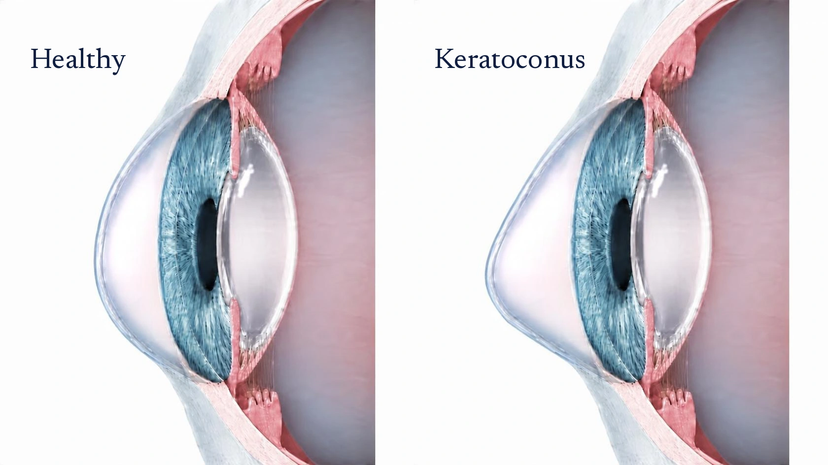
A healthy cornea is a smooth, evenly curved dome. In keratoconus, the cornea slowly thins and weakens, and the pressure inside the eye pushes it forward into a cone. The irregular shape scatters light, so vision becomes blurred and distorted.
It often begins in the teens or twenties and can progress for years. Common signs include frequent changes in glasses, rising astigmatism, glare and halos at night, and vision that glasses no longer fully correct. Eye rubbing, allergies and a family history all raise the risk.
How crosslinking works
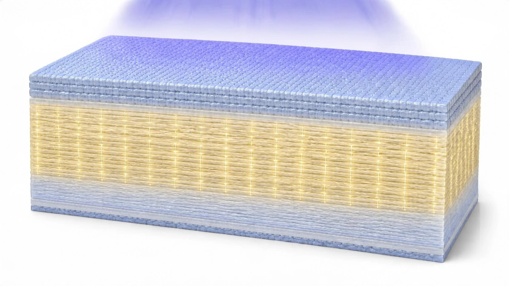
The cornea gets its strength from layers of collagen fibers. Crosslinking adds new bonds between those fibers, much like adding rungs to a ladder, so the cornea becomes stiffer and better able to hold its shape.
After numbing drops, the thin surface layer is usually removed so the medicine can soak in. Riboflavin (vitamin B2) drops are applied for a period of time, then a controlled ultraviolet light activates them. A soft bandage contact lens protects the eye while the surface heals.
What to expect
The first few days
The eye is often sore, watery and sensitive to light while the surface heals under the bandage lens. Drops help with comfort and protect against infection.
Vision
Vision can be hazy and fluctuate for several weeks to months as the cornea settles. Crosslinking is meant to stop the cone from worsening; it is not designed to remove the need for glasses or contacts.
Follow-up
Scans of the cornea’s shape are repeated over time to confirm it has stabilized. Avoiding eye rubbing helps protect the result.
Seeing clearly with keratoconus
In early keratoconus, glasses may be enough. As the cornea becomes more irregular, specialty contact lenses usually give the sharpest vision.
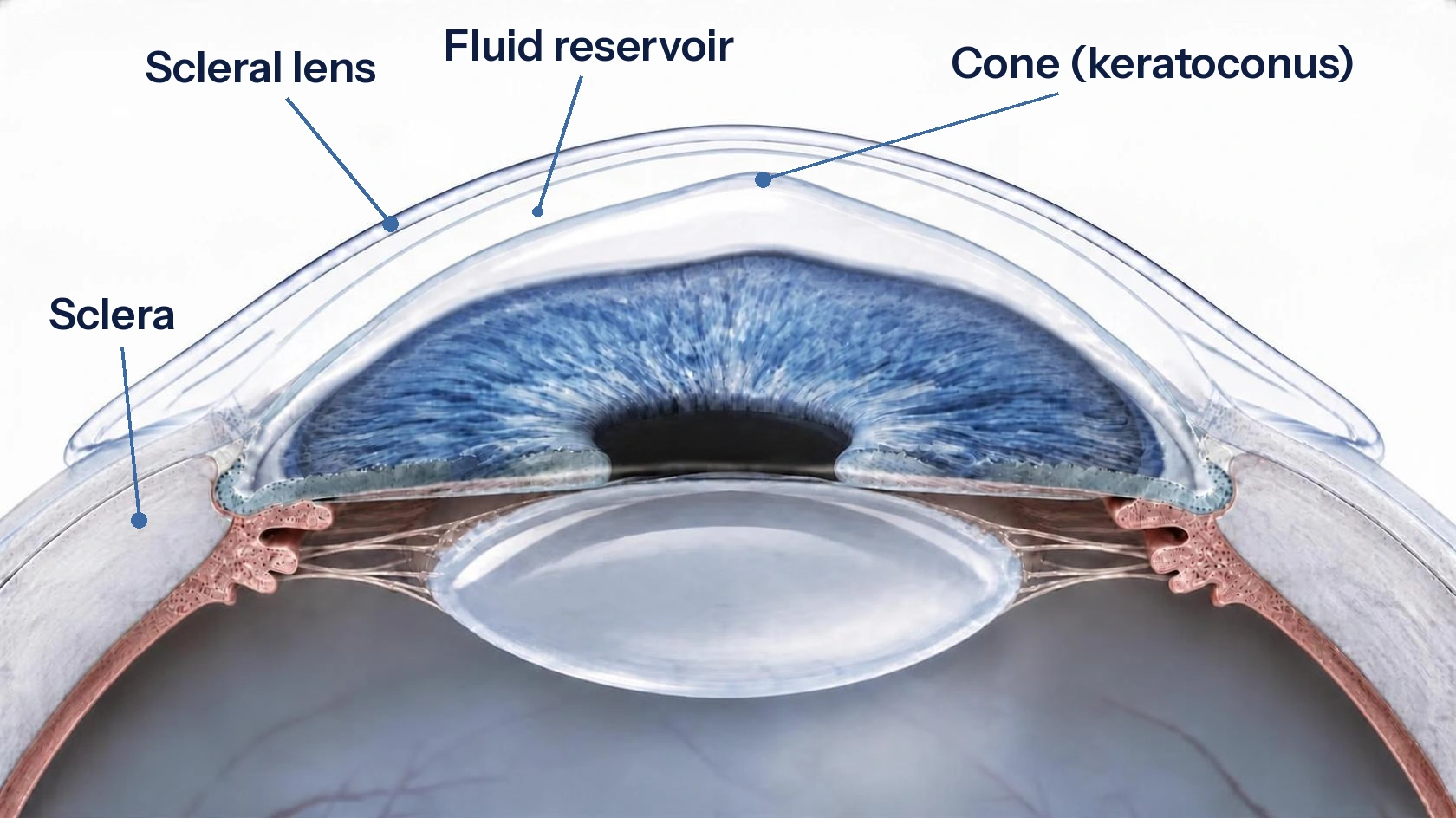
A scleral lens is a large, rigid lens that rests on the white of the eye and vaults completely over the cornea. The space underneath is filled with saline, which smooths over the irregular cone and gives light a clear, even surface to pass through. Scleral lenses are often comfortable, because they never touch the sensitive cornea.
If the cornea becomes too thin or scarred for lenses to help, a partial-thickness transplant (DALK) or a full-thickness transplant can replace the damaged tissue.
Every procedure has benefits and risks. Your surgeon will explain which options fit your eyes and why.