

Why pressure matters
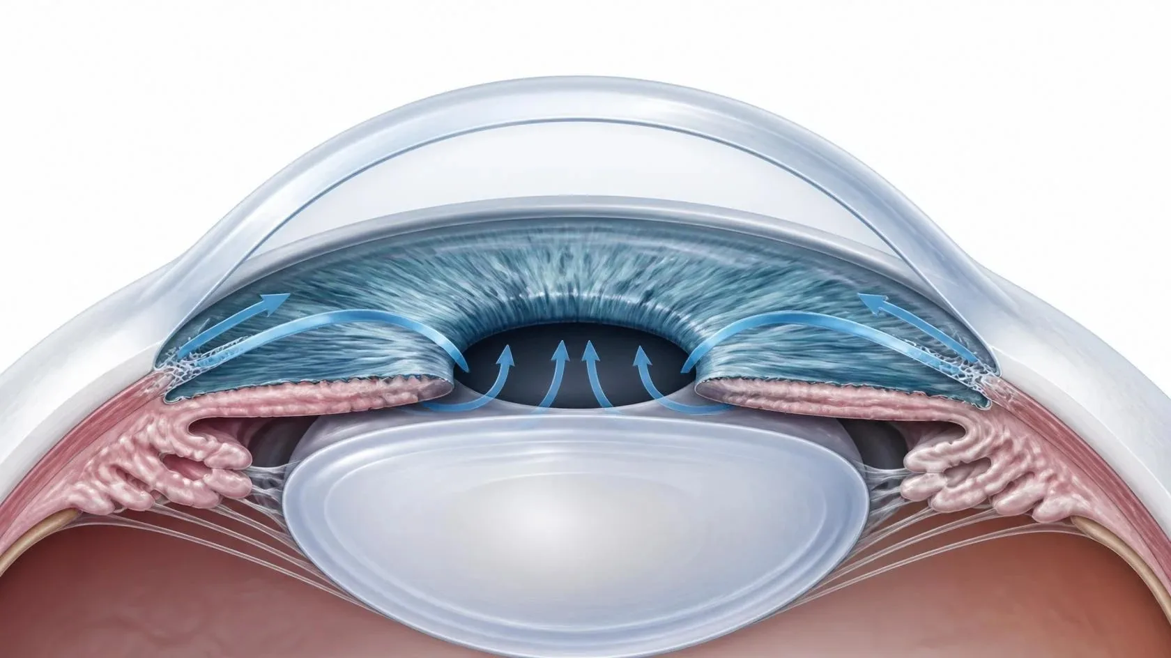
The front of the eye is filled with a clear fluid that is constantly made and drained. If the drain does not work well, pressure inside the eye can rise and, over time, damage the optic nerve.
Glaucoma can also occur with normal eye pressure, which is why the optic nerve itself is examined closely.
Two main types
Open-angle glaucoma
The most common type. The drain is open but works poorly, and pressure rises slowly. There is usually no pain, and side vision fades so gradually that it often goes unnoticed.
Angle-closure glaucoma
The iris blocks the drain. It can develop slowly, or suddenly as an emergency with eye pain, headache, blurred vision, halos and nausea.
How glaucoma is found
Because early glaucoma has no symptoms, regular eye exams are essential. Testing usually includes measuring eye pressure, examining the optic nerve, checking side vision with a visual field test, imaging the nerve fiber layer, and looking at the drainage angle.

 Healthy vision With glaucoma
Treatment
Vision lost to glaucoma cannot be restored, but lowering eye pressure can slow or stop further damage. Many people start with drops or SLT laser, and some need surgery to protect their sight.
Eye drops
Daily drops that reduce how much fluid the eye makes or help it drain.
Laser (SLT)
Selective laser trabeculoplasty, or SLT, is a quick in-office laser that helps the eye’s natural drain work better. It can be a first treatment instead of drops, or added to them, and can be repeated if needed. A different laser can make a small opening in the iris to relieve or prevent angle closure.
Surgical options, from gentle to advanced
When drops and laser aren’t enough, surgery can create a better way for fluid to leave the eye. The right choice depends on how advanced the glaucoma is, your eye pressure goal, and any previous surgery.
MIGS
Minimally invasive glaucoma surgery
Tiny devices or micro-procedures that improve the eye’s natural drainage system. MIGS is often performed at the same time as cataract surgery and usually means a quicker recovery. It is generally used for mild to moderate glaucoma.
XEN Gel Stent
A new drainage channel
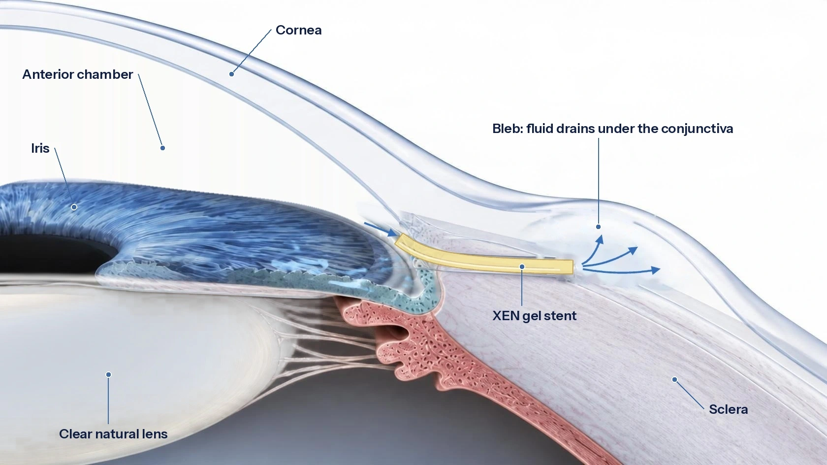
A soft, tiny tube about 6 mm long, placed through a small opening. It creates a new path for fluid to drain from inside the eye to a space beneath the eye’s thin outer covering. It can lower pressure more than MIGS, with a less invasive approach than traditional filtering surgery. See how XEN works
Glaucoma Drainage Tubes
Ahmed and Baerveldt implants
A small, flexible tube carries fluid from inside the eye to a plate placed on the eye’s outer wall, hidden under the eyelid. Tubes are often used for advanced or hard-to-control glaucoma, or after other surgeries.
Every surgery has benefits and risks. Your surgeon will explain which option fits your eyes and why.
The XEN Gel Stent
The XEN Gel Stent is a soft, flexible tube about 6 mm long, thinner than an eyelash.
Through a tiny opening, the stent is passed from inside the eye, through the drainage angle and the wall of the eye, so its far end rests beneath the conjunctiva, the thin clear covering over the white of the eye. Fluid then flows through the stent and collects in a small, shallow pocket called a bleb, where it is gently absorbed. This lowers eye pressure with a smaller incision and usually a quicker recovery than traditional filtering surgery.
XEN can be done on its own or together with cataract surgery.
Who is at higher risk?
- Age over 60
- A family history of glaucoma
- High eye pressure
- African, Hispanic or Asian heritage
- Diabetes, long-term steroid use or a past eye injury